


Because humans walk upright, the spine, as an axial structure, bears the main load. That is why degeneration and dystrophy are quite common consequences of human life. One of the most common diseases of the musculoskeletal system is osteoarthritis, which causes severe discomfort and can lead to disability. This article will discuss the most severe form of this pathology - common osteochondrosis.
General features
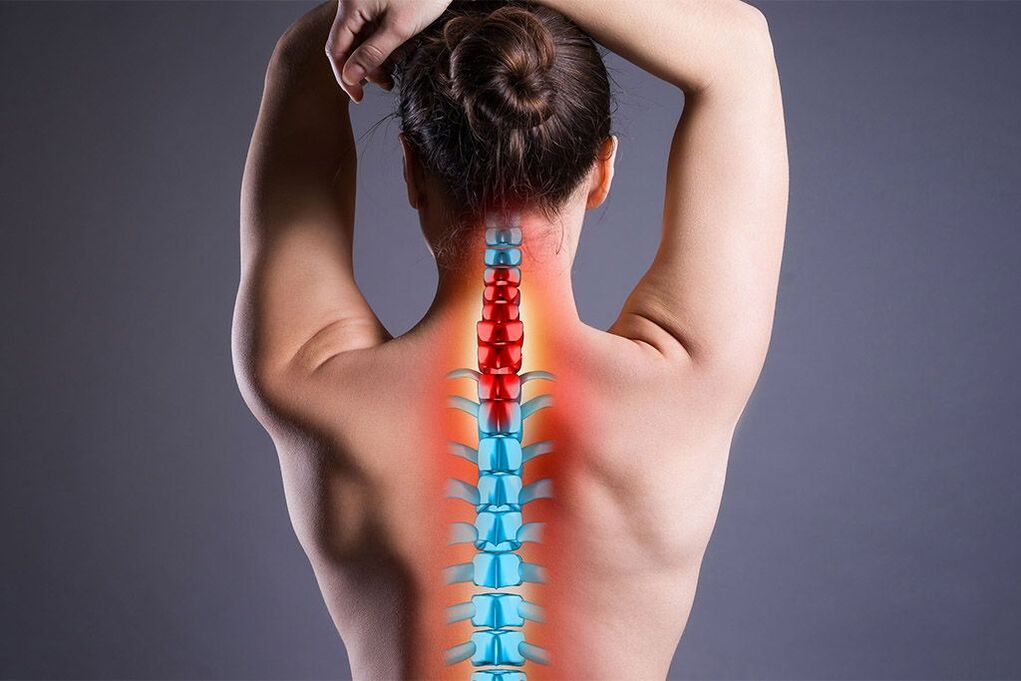
Osteoarthritis is a degenerative disease of the spine, most commonly affecting the thoracic, lumbar and cervical areas. This pathology has a direct correlation with age. The disease is much more common in people over 40 years old but has recently shown a trend in younger people. Ordinary osteochondronecrosis differs in that it affects multiple parts of one organ or several organs at the same time. Due to the increasing development of degenerative processes not only in bone tissue, but also in the ligamentous apparatus of the spine, the vertebrae become mobile and put pressure on the nerves and blood vessels. Symptoms of osteoarthritis are usually associated with this, but it is worth noting that the disease may be asymptomatic for some time.
Important! The pathology requires multidisciplinary management, as it affects not only the musculoskeletal system but also the nervous system as well as internal organs. In addition to the spine, the pathological process can also affect other parts of the skeleton.
Cause and pathogenesis
There are many reasons leading to widespread osteochondrosis. Some of them are associated with congenital bone defects, others are caused by insufficient load during vigorous activity. Particularly common factors that contribute to the development of the clinical picture are:
- injury;
- flat feet;
- Clubfoot - deformation of the foot (equinovarus, varus, valgus, depending on the position of the heel);
- work that involves lifting heavy objects;
- playing sports without warming up or warming up your muscles;
- work at low temperature.
Low temperature is considered an irritating factor, because cold temporarily changes the molecular structure of soft tissues, reduces the intensity of blood circulation, reduces the conductivity of nerve and metabolic impulses, and thereforereduces immune system activity. Other reasons disrupt the biomechanics of the spine and contribute to rapid disc wear.
Pain from widespread cartilage degeneration may be the result of bone spurs or disc deformity. The pain is often nerve root in nature, i. e. associated with compression of the posterior nerve root.
Common osteoarthritis easily mimics other diseases. When damaged in the chest area, pain appears in the heart area and is mistaken for an ischemic process, and with damage to the lumbar area - it is due to radiculitis.
Symptom
Clinical manifestations will depend on which organs are affected and what combination.
When the cervical spine is affected, the following characteristics are present:
- unstable blood pressure;
- headache;
- lack of coordination;
- pain in the hands;
- numbness in the upper body and arms.
For diseases in the chest area:
- intercostal neuralgia;
- stiffness in the arms and neck;
- dysfunction of internal organs.
If the lumbar region is affected:
- burn;
- urinary disorders;
- spasm;
- pain when walking.
Based on the above, it can be easily concluded that the pathology affects not only the spine and large joints, but also the autonomic nervous system. The second type is associated with disruptions in the functioning of internal organs. Normal multisegmental osteochondrosis can sometimes become more severe. In such cases, the manifestations are much more intense. With a combination of disorders of several organs, the symptoms will correspond.
symptoms
Osteoarthritis can be divided into moderate osteochondrosis, which is a natural process of wear and tear of the spine due to living activities, and severe osteochondrosis, which is often characterized by complications.
Moderate osteonecrosis can be easily treated with conservative treatment. And if it is impossible to completely stop the inevitable aging process, it is possible to slow it down significantly. Complications that severe osteonecrosis can lead to are as follows.
- Inflammation of the spine.
- Degenerative disc.
- Spinal stenosis.
Important! The intervertebral disc acts as a shock absorber and reduces friction between the vertebrae. Degeneration of these structures can lead to protrusion of the nucleus pulposus and disc herniation. Protrusion leads to compression of the roots and pain.
Spondylosis is the degeneration of the facet joints that connect adjacent vertebrae. In other words, such joints are called facet joints. When articular cartilage is damaged, painful contact occurs between the vertebrae. With degeneration of the facet joints, bone growths most often appear, leading to degenerative disease of the spine.
Stenosis is a narrowing (in this case of the spinal canal). Most often, stenosis is the result of pathologies such as intervertebral hernia or spondylosis. The bony growth and protrusion of the herniation compress the nerve roots at their entry and exit points.
The clinical picture of severe osteoarthritis is the result of complications:
- chronic pain in the spine;
- friction of bone surfaces;
- stiffness;
- sudden muscle weakness;
- decreased reflexes;
- tingling in the extremities;
- diffuse pain;
- sciatica symptoms.
Sciatica is caused by compression of the sciatic nerve.
Classify
There are four levels of osteoarthritis. Classification occurs on the basis of the collected history and with the help of instrumental diagnostic methods. The main criteria in this classification are pain and neurological symptoms.
- I degree - the pain is easily relieved with medication.
- Grade II - characterized by persistent pain and spinal deformity with moderate neurological symptoms.
- Grade III - systemic pain, significant neurological symptoms.
- Grade IV - constant pain, diverse neurological deficits. Disorders of nerve impulse transmission. Paralysis and paralysis.
In cases of widespread dysplastic osteochondrosis, the patient's disability status is determined. Depending on the general condition of the patient, the degree and intensity of development of the clinical picture, disabilities can be divided into three groups.
Types of defects in osteoarthritis.
| Group | Description |
|---|---|
| First group | The functions of the spine are lost. The patient is unable to move and take care of himself. |
| Second group | The patient can move around and perform minor tasks but has frequent exacerbations. This operation is contraindicated or useless for a number of reasons. Or surgery was performed but was ineffective. |
| Third group | The patient is capable of self-care. There are pain and vestibular symptoms, but the frequency of exacerbations is moderate and cyclical. |
Disability groups are classified by doctors based on a number of studies to evaluate working ability.
Diagnose
When you see your doctor, the diagnosis will include several components. First and foremost is the collection of history based on subjective information provided by the patient. Attention is paid to family history, because osteoarthritis has a genetic component. The specialist asks about the place of work, living conditions and the course of the disease, and the patient must describe exactly what is worrying him. The best results can be achieved if there is good feedback between patient and doctor.
The next method is objective research, carried out by experts themselves or using instrumental methods. The doctor checks the range of motion of the neck and limbs, which may be markedly reduced due to pain and stiffness. Using palpation, he recorded the degree of muscle spasm and the curvature of the spine. Attention is paid to neurological examination, thanks to which weakened reflexes can be detected. This symptom may be the result of nerve compression or damage.
Common methods of diagnosing cartilage degeneration include:
- X-ray of the entire spine in two projections.
- MRI to evaluate ligaments and nerve tissue.
- An electrophysiological study to examine nerve impulse conduction.
X-rays are effective in determining the presence of bone growths - bone spurs, spinal stenosis and the presence of other diseases that are a consequence of osteoarthritis, such as scoliosisliving.
Computed tomography may also be used in combination with MRI. Using a CT scan, you can determine the degree of compression of the nerve by the spur.
The diagnosis of widespread multisegmental osteochondrosis is made if other pathologies causing vertebral destruction (eg, tuberculosis) have been excluded and if several segments of one or more vertebral bodies are affected.
There are additional diagnostic methods. Including:
- Bone scan.
- CD.
- Myelogram.
Bone scans can detect conditions such as osteoarthritis, fractures or infections. This method is radionuclide, suitable for differential diagnosis and identification of possible complications.
During a discogram, a contrast agent is injected into the nucleus pulposus of the disc. This method is effective in determining the presence of disc herniation.
Myelogram is also a contrast study method. Contrast material is injected into the spinal canal and images are recorded using X-rays or CT. Using this method, you can determine the condition of the spinal cord, the presence of narrowing and compression points.
Treatment
Treatment is based on the following mechanisms.
- Slows down the degenerative process by improving the supply of nutrients to the structures of the musculoskeletal system.
- Stabilize the spine.
- Removes pinched nerve fibers.
- Reduce symptoms.
The following drugs are used for drug treatment:
- Nonsteroidal anti-inflammatory drugs help reduce inflammation and pain;
- Anilides reduce pain in the early stages;
- local analgesics in ointment form;
- muscle relaxants to reduce muscle spasms and increase range of motion;
- Vitamin B to improve nerve tissue conductance;
- chondroprotector, reduces the rate of progression of degenerative processes by integrating active substances (chondoitin sulfate and glucosamine) into chondrocytes. As a result, metabolism is normalized and clinical manifestations are reduced. The drug has been used for a long time and requires special advice during pregnancy, lactation and in the presence of diseases of the gastrointestinal tract. An absolute contraindication is phenylketonuria;
- Antispasmodic drugs reduce smooth muscle spasms and thereby reduce the manifestations of osteoarthritis in internal organs;
- Antioxidants;
- Antidepressants to eliminate the psychological component of this disease. They interfere with the transmission of nerve impulses from the central nervous system to the brain. Promotes endorphin production and helps solve the problem of chronic insomnia caused by constant pain.
- neuropathogenic agents to eliminate damage to nerve endings.
- opiates to relieve intolerable pain and the ineffectiveness of other analgesics.
The following are used as invasive medical procedures:
- steroid injection into the epidural space. Steroids are powerful anti-inflammatory drugs. They reduce nerve root inflammation, helping to relieve pain caused by radiculopathy. Complicated procedures. Ask for a qualified professional;
- injected into the facet joint. The injection has local anesthetic and pain-relieving effects.
It's important to know! Medication is not intended to cure the disease - there is no medication that can completely eliminate osteonecrosis, a chronic disease. Medicines are prescribed only to relieve symptoms.
The medicine is prescribed by the attending physician. Patients are informed about the possible side effects of each drug and then decide for themselves which treatment to choose.
For symptoms that make you suspect osteoarthritis, contact a spine specialist, orthopedist, and neurologist. High-quality medical care will include close collaboration between these professionals with each other and with patients.
Physical therapy
Physiotherapy is used as a set of auxiliary therapeutic measures to improve blood circulation and metabolism in affected tissue. For widespread osteonecrosis, the following methods are used.
- Electrophoresis (based on the movement of colloidal particles under the influence of an external electric field).
- Phonophoresis (combination of ultrasound and medication).
- Magnetic field therapy (using a static magnetic field).
- UHF therapy (ultra high frequency therapy).
- Electromyostimulation (stimulation of nerves and muscles).
- Acupuncture (acupuncture).
- Exposure to laser beams.
In addition to physiotherapy, manual therapy and physiotherapy are also actively used. Professional massage can lead to lasting relief. Exercise therapy should not be performed during periods of exacerbation of the disease, as this can lead to complications. During remission, moderate physical activity will maintain muscle tone and thus the spine. Exercises are performed under the supervision of an instructor and prescribed by the attending physician.
During an exacerbation, you cannot warm the spine but can wear a corset but only for a few hours. In other cases, wearing a corset for more than a few hours is not optimal because it can lead to muscle atrophy.
Manual therapy can increase the release of compressed nerves and reduce neurological symptoms. Alternative methods are leeching and vacuum massage. These methods are aimed at improving blood circulation in the affected area. Hygienic spa treatment is very helpful. Special priority is given to return home procedures.
Surgery
When treating osteoarthritis, experts are more willing to use conservative therapy, however, to get the desired effect, a lot of time, patience and careful compliance with the patient's recommendations are required. . If conservative treatment is not effective, you should use invasive methods. Usually the operations are palliative. This means that surgery will only be performed to relieve symptoms and improve quality of life moderately, not to completely cure it (it is fair to say that conservative treatment also does not lead to elimination). completely eliminate the disease, but rather the patient's ability to absorb it). Switching to non-invasive treatment is a sign of good prognosis).
There are two types of surgery: decompression and stabilization. The first is aimed at relieving nerve compression, and the second is aimed at stabilizing the spine. The following activities are classified as decompression activities.
- Facetectomy – removal of the facet joints to relieve compression.
- A foraminotomy is an increase in the lumen of the spinal canal, which has been narrowed by osteoporosis.
- Surgical excision of the posterior portion of the vertebrae may result in deformity due to osteonecrosis.
- Laminectomy - removing a piece of the back part of the vertebra to widen the spinal canal.
These surgeries require a posterior approach, but in the case of intervertebral hernia, the surgical approach will be anterior.
Surgical decompression with an anterior approach is as follows.
- Discectomy - removal of a disc.
- Vertebrectomy – removal of the entire vertebral body with adjacent discs.
Stable operation includes:
- Vertebral fusion is a method of fusing the vertebrae.
- Artificial disc.
The need for functional stabilization arises after discectomy.
Surgery is rarely indicated because of the risk of developing serious complications.
Complications include:
- pain recurrence;
- wrong combination;
- infection;
- phlebitis in the limbs;
- violation of urination behavior;
- pain due to grafting;
- failure of embedded screws.
The postoperative period lasts several months. The stitches will heal 2 weeks after surgery. If complications are detected, you should consult your doctor immediately.
After surgery, a rehabilitation course is carried out to accelerate wound healing and restore full working ability.
General recommendations
Proper nutrition helps prevent recurrence of exacerbations of common osteoarthritis. Proper nutrition is primarily necessary to maintain a stable body weight, as excess weight puts additional stress on the spine. In this case, the diet must be complete, fortified and rich in calcium, magnesium and potassium. Coffee consumption should be limited because it will leach calcium from the body. It is very useful to visit the swimming pool. You should avoid staying in one position all the time.
If there are frequent exacerbations and lack of discipline to strictly adhere to recommendations, it is best to undergo a full course of treatment in the hospital under the supervision of a doctor.
You cannot take the medicine yourself.
Let's summarize
Typically, extensive osteochondronecrosis develops after incomplete resolution of "isolated" osteochondronecrosis. This fact shows that if any unpleasant sensations occur, you should immediately contact your doctor, and not hope that the pain will go away on its own. In this case, it will be much easier to prevent the development of other pathologies, and it will be much easier to cure them at the root.